Retinal and vitreous surgeries are known to be among the most delicate and sensitive eye surgeries, as they target the most critical parts of the eye, especially the visual cortex, which is responsible for sharp vision.
Accordingly, this procedure requires strong medical expertise and very high surgical skill, which are found in Dr. Sharif Mumtaz Hijazi, a consultant in ophthalmology and eye surgery.
Retinal and vitreous surgeries
Before discussing retinal and vitreous surgeries, it is necessary to define these highly delicate parts of the eye.
The term retina refers to the very thin tissue layer located on the back endothelial wall of the eye. In the center of this layer is the retinal macula, also known as the visual cortex, which is directly connected to the optic nerve.
As for the vitreous body, it is a transparent, gelatinous body made up of a high percentage of fluids and proteins. It is located in the inner chamber of the eye and gives the eye its spherical shape.
The most important diagnostic tests before retinal and vitreous surgeries
At Dr. Sherif Mumtaz Hijazi’s center, the medical team works to fully assess the patient’s condition before taking any action, no matter how simple. This, of course, applies to retinal and vitreous surgeries, which are the most cautious, as a series of diagnostic tests are performed beforehand to guide the patient to the appropriate treatment based on their condition. These tests include:
- Optical coherence tomography (OCT): This involves imaging all tissue layers of the retina and its blood vessels without the need to inject a colored or radioactive substance to assess the health of the retinal layers and blood vessels.
- Fluorescein angiography: Fluorescein dye is used to reveal any blockage, clot, or abnormality in the blood vessels within the retina after a certain period of time following injection and undergoing specialized imaging.
- Retinal electrophysiology test: In this test, the electrical response of retinal activity to light rays is measured.
Causes of retinal and vitreous diseases
Aside from the procedures and types of retinal and vitreous surgeries, there are many factors that cause retinal and vitreous disease and necessitate surgical intervention, including:
- Having diabetes, which causes episodes of high blood sugar that damage the blood vessels in the retina and cause blood and fluids to leak from them into the retina and vitreous body.
- Chronic high blood pressure that affects the blood vessels that nourish the retina.
- Direct injuries to the eye and severe trauma that cause damage to the retina.
- Genetic factors that interfere with retinal health, such as Stargardt genetic disease and Usher syndrome.
- Smoking and alcohol addiction affect the blood vessels in the retina and reduce the amount of oxygen reaching them.
- With advancing age, the proportion of fluids in the vitreous body increases and it becomes more fluid.
- Severe myopia increases the likelihood of premature vitreous detachment.
- Eye surgeries that affect the integrity of the retina and cause problems in it.
- Having glaucoma, or blue water, which raises eye pressure and causes damage to the retina.
Retinal and vitreous diseases
Retinal and vitreous surgeries are used for dozens of different diseases, and often a single surgery or treatment technique is extensive, addressing several diseases simultaneously. Among the most important of these diseases affecting the retina and vitreous body of the eye are:
- Congenital retinal atrophy.
- Diabetic retinopathy.
- Retinal detachment.
- Retinal tear.
- Diabetic macular edema.
- Age-related macular degeneration.
- Blockage of retinal veins and arteries.
- Central retinopathy.
- Hereditary retinal degeneration.
- Retinitis.
- Retinal tumors.
Symptoms of retinal and vitreous problems
When diagnosing any retinal problems or diseases, a complete and detailed medical history of the patient will be taken. Among the most important parts of the patient’s clinical history are the symptoms and signs they are experiencing, the timing of their onset, their severity and frequency, and other details regarding them. Some of the most important signs and symptoms of damage to the retina and vitreous body of the eye are:
- Increased appearance of floaters in the eye such as black spots, zigzag lines, and dots that resemble a fly.
- Seeing flashes of light, similar to lightning, in the peripheral vision.
- Seeing a shadow or moving image within the field of vision is evidence of retinal detachment.
- Vision loss gradually from the periphery to the center.
- Continuously and increasingly blurred vision.
- Seeing columns as zigzag and straight lines as curved, and clear visual disturbances.
The most important retinal and vitreous surgeries
After diagnosing the retinal condition and referring the patient for a necessary retinal and vitreous surgery, Dr. Sharif discusses all aspects of the surgery and its outcomes with the patient to ensure they understand the expected results, the path to achieving them, and the timeframe for reaching them. These available surgeries include:
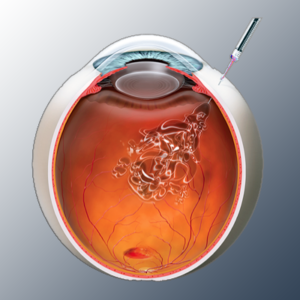
Intravitreal injection
In this procedure, therapeutic substances are injected directly into the vitreous body. These drugs vary depending on the patient’s condition. For example,
Vitrectomy surgery
In the event of a problem with the retina, or in cases of retinal detachment or vitreous hemorrhage, surgery is performed to completely remove the vitreous body from the eye cavity and replace it with a bubble of gas, silicone oil, or a special solution.
Retinal grafting procedure
In this treatment technique, a bubble of gas or air is injected into the middle of the vitreous body, where this bubble takes its proper place and then pushes the damaged retinal layers towards the wall of the eye to stop fluid leakage to the back of the retina.
Photoretinal therapy
Through an advanced mechanism, a drug called vertebrophen is injected into a vein in the arm. This drug then automatically travels to the blood vessels within the eye over a certain period. After that, special laser beams are directed at the blood vessels in the retina in order to stimulate the activation of vertebrophen, which works to close the blood vessels in the retina.
Laser photocoagulation
In the early stages or before the onset of retinal detachment or diabetic retinopathy, photocoagulation can be performed using high-energy laser beams that cause a direct burn to the retina to reduce retinal detachment or stop a minor tear at a certain point.
Gene therapy for the retina
It is one of the most advanced techniques in retinal and vitreous surgery, where genetic factors are used to repair the defect.
Here, the patient’s damaged genes are replaced with healthy, problem-free genes through local injection into the retina. This treatment is still in the experimental stage, but its results are promising and could open up vast possibilities for treating patients.
Drug therapies that have recently been licensed by the U.S. Food and Drug Administration may be used for this purpose.
Steps of vitreous surgery in the eye
Vitreous surgery is one of the most prominent retinal and vitreous surgeries that aims to remove the vitreous body to treat problems of retinal detachment and bleeding in the vitreous body, in addition to retinal inflammation and holes. This surgery is performed in the following steps:
- The patient is anesthetized locally using anesthetic eye drops.
- Making precise surgical incisions on the outer surface of the eye, reaching down to the vitreous body.
- Removal of the vitreous body from the eye, which is damaged and sometimes tinged with blood.
- Repairing retinal defects and problems in any suitable way.
- Filling the vitreous body space with injections of gas, silicone oil, or a customized saline solution.
- Closing surgical incisions on the surface of the eye with surgical sutures or other techniques.
Steps of retinal surgery in the eye
The range of diseases and problems affecting the retina is much wider than that affecting the vitreous humor of the eye. These include retinal tears, retinal detachment, macular edema, and many others. Some of the most common treatments for these conditions are:
·Air retinal fixation
In the case of retinal detachment, the condition can be treated by injecting a bubble of gas into the vitreous cavity of the eyeball.
This gas bubble works by putting pressure on the detached retina in a satisfactory way, and thus the retina returns to its correct position.
After this procedure, the patient must follow many instructions and maintain a specific head position when sleeping and sitting to keep the gas bubble in its correct position.
Pneumatic retinal fixation may be used in conjunction with photocoagulation or freezing to achieve optimal results.
Laser photocoagulation
The most suitable treatment for retinal tears and holes is based on the principle of using laser beams to make small burns around the site of the retinal tear. When the retina heals, scar tissue will form automatically. This repaired tissue will, in turn, close the retina and all the holes in it.
This procedure is adopted as a preventive measure in case a minor tear in the retina is detected before retinal detachment occurs.
·Cooling
This technique treats retinal tears using the same mechanism as laser treatment, but with extremely high temperatures.
A freezing probe is applied to the outer surface of the eye in order to freeze the area surrounding the tear site in the retina, thereby creating scar tissue to repair the retinal tissue.
Advantages of retinal and vitreous surgeries
There are many advantages and benefits to undergoing retinal and vitreous surgeries. These surgeries are suitable for patients of all ages and boast very high success rates. Among the most important of these advantages are:
- High effectiveness in treating retinal and vitreous diseases, no matter how complex they may be.
- It significantly improves vision quality and corrects vision defects to a remarkable degree.
- Protecting the eyes from complications of retinal diseases and reducing the likelihood of vision loss resulting from them.
- Performing surgeries using modern and advanced techniques with very rare risks and side effects.
Defects in retinal and vitreous surgeries
Like any other treatment or eye surgery, there are some drawbacks and disadvantages that cannot be ignored. However, at Dr. Sherif Mumtaz Hegazy’s center, we adhere to the highest quality standards to achieve the lowest possible rates of potential complications, in accordance with the high quality of the treatment procedures adopted at the center. Nevertheless, the potential drawbacks of these surgeries include:
- Bacterial or viral infections resulting from contamination of surgical instruments or improper eye care after the operation.
- Bleeding inside the eye during or immediately after the procedure as a result of damage to the blood vessels in it.
- The condition of cataracts, if present before, deteriorates very rapidly after surgery.
- Elevated eye pressure and severe glaucoma require immediate treatment to prevent affecting the results of surgery.
- Weakened central vision and seeing blind spots in the center of the visual field, along with seeing increasing amounts of floaters.
- There is a possibility of relapse of retinal detachment surgery despite the surgery being successful the first time.
- Scar tissue and fibrous tissue form on the retina, especially in complicated cases of retinal diseases.
- Vision loss occurs in rare cases, especially in the very late stages of retinal diseases.
Key guidelines after retinal and vitreous surgeries
Any retinal or vitreous surgery, no matter how simple, requires meticulous post-operative care to ensure a smooth recovery without complications and to achieve complete healing as quickly as possible. Among the most important medical guidelines and advice that Dr. Sherif Mumtaz Hegazi gives to his patients who have undergone retinal or vitreous surgery are:
- Recognizing the normal side effects after surgery, such as eye redness, blurred vision, moderate pain, and other symptoms that are part of the recovery phase and nothing to fear, and clearly distinguishing them from complications of the operation.
- Regular follow-up at Dr. Sherif Mumtaz Hegazy’s clinic and checking the condition of the eye, the health of the retina and the vitreous body after the operation.
- Avoid strenuous physical activities such as lifting heavy weights, doing strenuous exercise, or even flying.
- Avoid strenuous physical activities such as lifting heavy weights, doing strenuous exercise, or even flying, as this may cause a change in air pressure and thus affect the gas bubble injected into the eye.
- Avoid rubbing or scratching your eyes to protect your retina from exposure to high pressure or unintentional external trauma.
- Practice the eye exercises prescribed by the doctor if needed, the most important of which is not to move the eye too quickly.
- Completely refrain from swimming, exposing the eye to water, washing it, or applying cosmetic products to it until it is fully healed.
- Avoid excessive use of electronic devices such as televisions, mobile phones, and computers, as they cause eye strain.
Best eye doctor in Egypt
When it comes to retinal and vitreous surgeries, not just any ophthalmologist can perform these procedures. The surgeon must be highly skilled, with deep expertise, advanced surgical abilities, and exceptional precision. Among the elite Egyptian ophthalmologists renowned for retinal and vitreous surgeries is Dr. Sherif Momtaz Hegazy, who has spent decades refining unparalleled surgical skills and medical expertise across the Middle East, earning a distinguished reputation in the international medical community.
This is because Dr. Sherif worked in dozens of prestigious jobs and positions in several countries around the world after graduating from medical school and obtaining a master’s degree in ophthalmology from Cairo University in Egypt, which enhanced his reputation and medical expertise on both the academic and professional levels. Among the most important of these jobs are:
- Teaching at the Research Institute of Ophthalmology in Egypt.
- Joining the Royal College of Surgeons in the United Kingdom.
- Obtain an international scholarship for excellence in the World Council of Ophthalmology.
- Fellowship at the University of Tübingen in Germany, specializing in retinal and vitreous surgery.
- Accreditation as a trainer at the Eye Diseases Research Institute in Egypt.
- International examiner for the Fellowship of the Royal College of Surgeons in the specialty of ophthalmology in Britain.
- Scientific researcher and international lecturer at the most prestigious international medical conferences and institutions.
All these achievements are but a drop in the ocean of perseverance, striving, and reaching the top through diligent work and escalating experience that Dr. Sherif Mumtaz Hegazy has honed over many years.
This journey has been distinguished by bringing together the essence of long experience in the field of ophthalmology and eye surgery at the Dr. Sherif Mumtaz Hegazi Center in Egypt, which has been equipped with the best means and the latest diagnostic and therapeutic technologies, from microscopes and imaging devices to advanced LASIK releases and integrated equipment to provide the best medical services to patients and treat all vision problems accurately and with the highest possible success rate.